
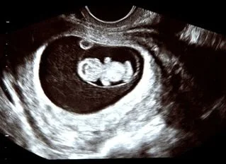

Anomaly Scans
Worried about your Anomaly scan results chart? Get a second opinion from the best fetal medicine specialist in Chennai, Dr. Raghav Arora

Experience
With over 15 years of experience,, Dr. Raghav Arora has performed over 1,50,000 successful scans.
Same Day Report
We understand your nervousness after a scan, so we provide same-day reports to ease your mind and save your time.
Got a question?
Frequently Asked Questions
Which week is best for an anomaly scan?
The best week for anomaly scan is usually between 18 and 24 weeks of pregnancy.
This particular period of gestation is the best time frame to visualize the baby’s structures and anatomy to provide information on the risk of birth defects or abnormalities.
During the 20 week scan heart abnormalities like holes larger than 5mm or any malformation in the arteries can be clearly seen.
Will the Anomaly Scan Provide 100% Confirmation Of The Structural Development?
The answer is NO.
However, the anomaly scan is highly accurate, yet it cannot detect 100% of structural abnormalities.
While most babies are born healthy, only a small percentage of them stand the risk of developing complications.
An anomaly scan cannot detect such complications that evolve during the later phases of gestation.
What if there are signs of a problem in an Anomaly Scan?
the defect revealed will be studied in detail. Following that, your doctor offers you genetic counseling.
How to Prepare for an Anomaly Scan?
However, a full bladder is not necessary for an anomaly scan. The gynecologist may ask the expectant mother to drink enough water to fill her bladder in certain conditions.
Thus, it helps lift the uterus and gives a clear picture of the fetus. Likewise, it is necessary to wear comfortable or loose-fitting clothes.
Do not apply oil or lotion to the belly.
Can I Eat Before Anomaly Scan?
Yes, you can eat before the anomaly scan. However, your gynecologist may ask you to avoid eating gas-producing foods, like broccoli or beans, as they may cause discomfort during the scan.
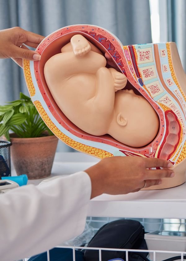
What is the Anomaly Scan Procedure?
An anomaly Scan Procedure is : the pregnant mother is asked to lie down comfortably on the examination table in the scanning station.
A thick layer of water-based gel is applied to the abdomen area to ensure that the probe can glide smoothly on the abdomen and provide good-quality images.
The sonographers use high-tech scanning equipment that emits high-frequency sound waves to produce quality images of the baby for accurate results.
These results will then be analyzed by the fetal medicine specialist.
How to Change a Baby’s Position for an Anomaly Scan?
However, it is quite similar to an NT scan. Your gynecologist might ask the expectant mother to try again after a break.
A sugary drink, chocolate, sweets, and a gentle movement may help the
What is a normal anomaly scan report?
A normal 20 week anomaly scan report specifies no structural abnormalities or birth defects that are detected during the examination.
Limitations of the anomaly scan
- Though the anomaly scan detects the abnormalities, your doctor cannot identify the reason behind malformation. In such cases, either a repeat test or genital counseling is recommended.
- Certain abnormalities like major cardiac issues, bowel obstructions, congenital diaphragmatic, hernia, hydrocephalus, microcephaly, club foot, kidney or intestinal obstruction may evolve during the later phase of pregnancy. The evolving variations cannot be diagnosed in an anomaly scan. Your fetal expert may recommend a repeat scan between 24 and 25 weeks of gestation in such situations.
- Some findings may not be anomalies but require scan follow-ups, biochemical testing or invasive testing. Some findings may be transient and may change with time
- Specialized Fetal Echo (heart study) or extended neurosonography (brain study) will be done if the referred doctor prescribes the same. Even if the brain may look normal on ultrasound examination, its function cannot be evaluated.
- Some anomalies like soft tissue fusion of fingers/toes, absence of anal opening or absence of auditory opening may not be recognizable by ultrasonography.
- Cleft lip found deep inside the mouth is a very challenging factor to be diagnosed with an ultrasound scan.
- Other factors such as the patient’s build, scars from a previous operation and the way the baby is positioned may limit the diagnostic ability of this test.
- Several factors like gestational age at which scan is done, the fetal position at the time of the scan, maternal body habitus, liquor volume, and shadows from fetal parts may restrict/limit the visibility of this scan.
- Ultrasound markers have only 50 to 70% capability to diagnose chromosomal anomalies. Consequently, invasive procedures for identifying Down’s syndrome may be the best option. Your fetal expert can advise you on these procedures.
- Multiple gestations (twin/triplet pregnancies etc.) may cause difficulties in ultrasound examination due to fetal position and overlap.
- Even specialized fetal echocardiography (heart study) cannot pick certain cardiac anomalies such as Secundum ASD, small VSD, PDA, etc. A heart hole lesser than 5mm is also hard to diagnose in an anomaly scan.
- Obesity is a special challenge for ultrasonography. The fat in the mother’s abdominal wall absorbs the ultrasound energy, thus degrading the images and making the diagnosis very difficult.
- 3D/4D ultrasound is used only to assess certain abnormalities and is not a routine.
- The study of the genital organs is prohibited under the PC & PNDT Act. Hence detection of abnormalities of genital organs is not feasible.